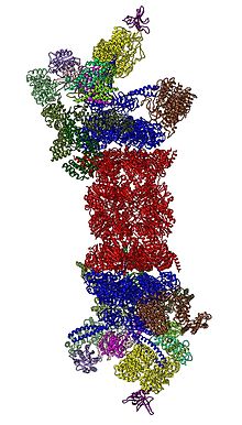
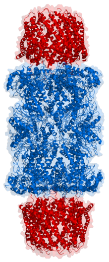
Proteasome / Proteasome Structure And Function A Structures Pdb 4r3o And Download Scientific Diagram
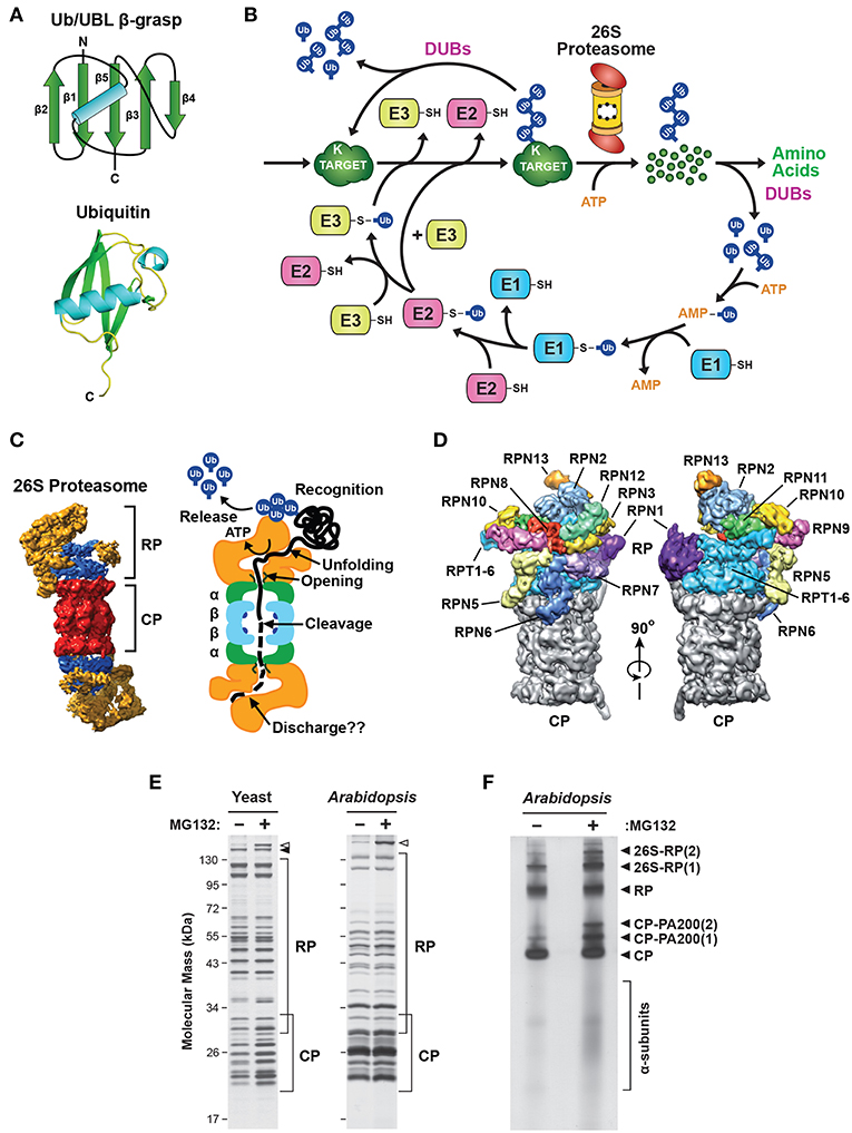
In this review we systematize current data. Upon entering the proteasome channel the polypeptide chain of the protein unfolds and stretches along it being hydrolyzed to short peptides.
Proteasome Wikipedia
The proteasome is a multisubunit enzyme complex that plays a central role in the regulation of proteins that control cell-cycle progression and apoptosis and has therefore become an important target for anticancer therapy.

Proteasome. Immunoproteasomes are induced by interferon gamma but also by other proinflammatory cytokines and oxidative stress which in the cell triggers the transcription of three catalytic subunits that do not occur. Diese Strukturen sind wichtig weil sie helfen diese beschädigten oder nicht benötigten Proteine loszuwerden bevor sie andere nahe gelegene. The luminescent assay format avoids inherent fluorescent background signals.
The Proteasome-Glo 3-Substrate System consists of three homogeneous bioluminescent assays in an enzyme-based format each of these three assays also is available separately. If it is to be used on a regular basis it is recommended that the material should be stored between 0C-4C during the duration of use. November 2011 von Herrn PD Dr.
Proteasome Activity Assay Kit ab107921 takes advantage of the chymotrypsin-like activity using an AMC-tagged peptide substrate Proteasome Substrate Succ-LLVY-AMC in DMSO which releases free highly fluorescent AMC ExEm 350440 nm in the presence of proteolytic activity. Diese Dissertation wurde im Sinne von 7 der Promotionsordnung vom 28. The kit also includes a positive control Jurkat Cell lysate with significant proteasome activity and a specific.
Proteasomes form the major non-lysosomal ATP-dependent protein degradation systems that targets intracellular polyubiquitinated proteins derived from self-structures or foreign structures for proteolytic degradation which includes the removal of misfolded and immature proteins and the production of peptides for presentation by MHC class I molecules. In human platelets. Of the proteasome.
The Proteasome-Glo 3-Substrate System consists of three homogeneous bioluminescent assays in an enzyme-based format each of these three assays also is available separately. However the degradation rate of proteins is also affected by the capacity of proteasomes to recognize and degrade these substrate proteins. This capacity is regulated by a variety of proteasome modulations including 1 changes in.
This process has been named Ub-dependent protein degradation. Proteasome-Glo Cell-Based Assays provide luminogenic proteasome substrates in buffers optimized for cell permeabilization proteasome activity and luciferase activity. Watch our science slam on proteasome function in response to cigarette smoke.
The simplified method minimizes handling steps and makes the assays amenable to automation. The simplified method minimizes handling steps and makes the assays amenable to automation. For longer term storage the.
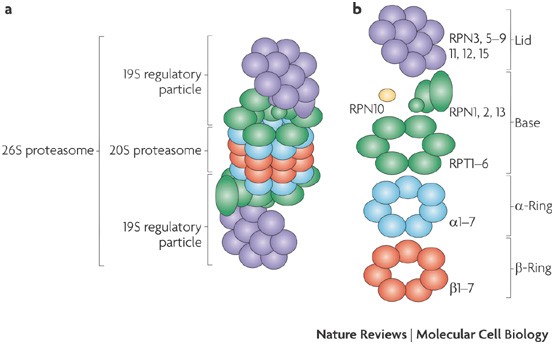
An immunoproteasome is a type of proteasome that degrades ubiquitin-labeled proteins found in the cytoplasmIn general proteasomes consist of a regulatory and a catalytic part. Our lab investigates the role of proteasomal protein degradation in chronic lung diseases. Proteasome sind große Proteinstrukturen mit mehreren Untereinheiten die zusammenkommen verschiedene Proteine abzubauen die entweder beschädigt sind Abfallprodukte enthalten oder auf andere Weise nicht benötigt werden.
Dictcc Übersetzungen für proteasome im Englisch-Deutsch-Wörterbuch mit echten Sprachaufnahmen Illustrationen Beugungsformen. Björn Krämer betreut und von Herrn Prof. An animation on Proteasomes.
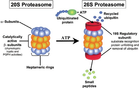
It has been demonstrated that 20S proteasome undergoes only slow loss of activity when stored at 4C for up to six months. Ubiquitin per se does not get into the proteasome but after destruction of the labeled molecule it is released and labels another molecule. The proteasome is responsible for the degradation of the majority of intracellular proteins which are often targeted for degradation via polyubiquitination.
The luminescent assay format avoids inherent fluorescent background signals. Addition of the Proteasome-Glo Cell-Based Reagent in an add-mix-measure format results in proteasome cleavage of the substrate and rapid generation of a luminescent signal produced by the luciferase reaction. 20S Proteasome remains relatively stable and active so long as repetitive freeze-thaw cycles are avoided.
Stefan Zahler von der Fakultät für Chemie und Pharmazie vertreten. Proper proteasome function is thus essential for numerous cellular processes such as protein turnover and quality control cell growth and cell signaling antigen presentation and immune response. Before a protein is degraded it is first flagged for destruction by the ubiquitin conjugation system which ultimately results in the attachment of a polyubiquitin chain.
Diese Dissertation wurde.
Proteasome Wikipedia

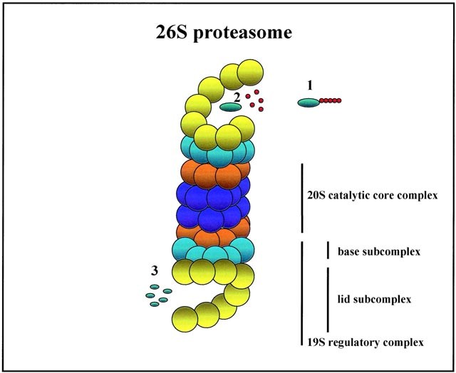
Schematic Representation Of Proteasome 26s Proteasome 26s Named Due Download Scientific Diagram
Role And Function Of The 26s Proteasome In Proliferation And Apoptosis Laboratory Investigation
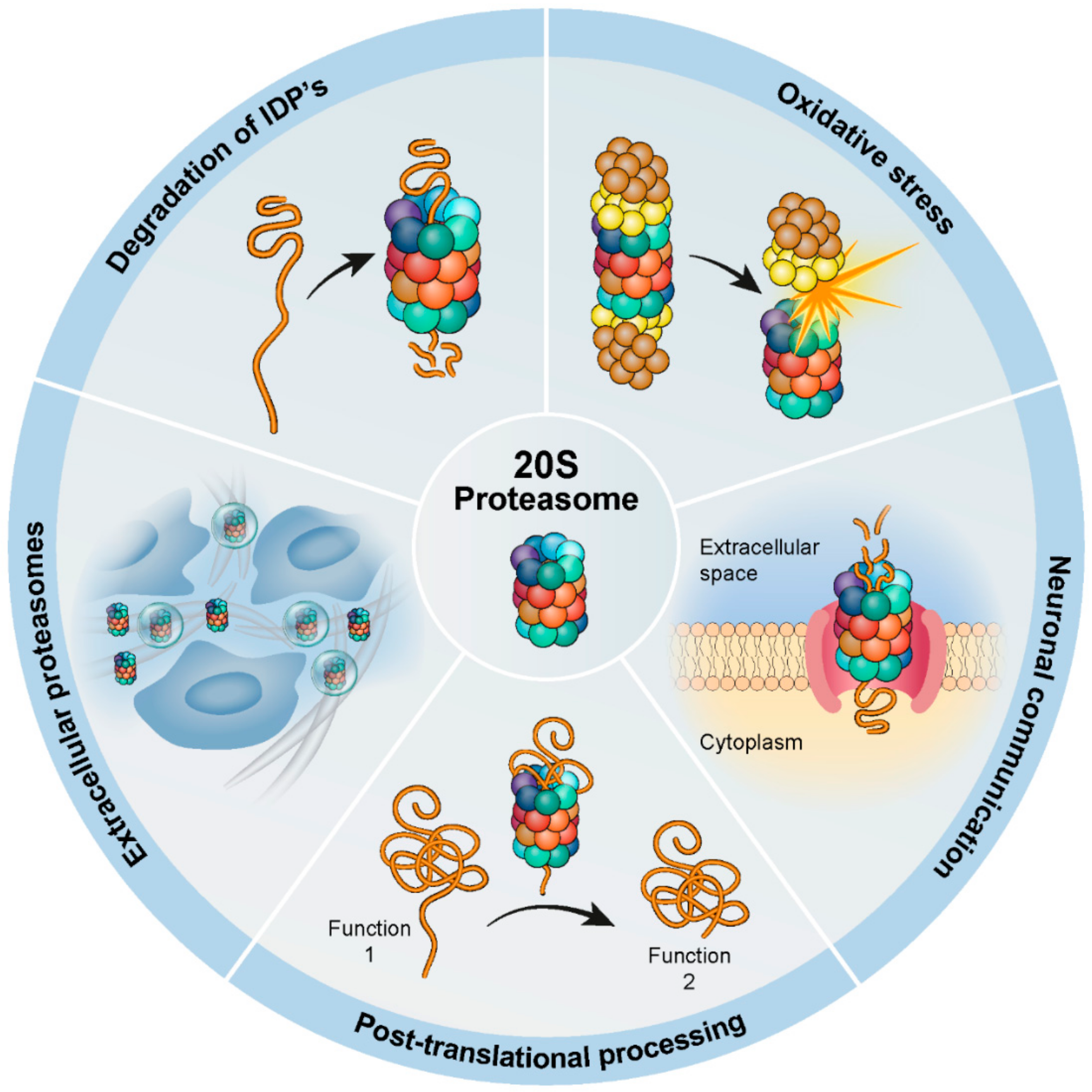
Biomolecules Free Full Text The Contribution Of The 20s Proteasome To Proteostasis Html
Kegg Pathway Hsa03050
Frontiers Dynamic Regulation Of The 26s Proteasome From Synthesis To Degradation Molecular Biosciences

Pdf Regulation Of Protein Degradation By Proteasomes In Cancer Semantic Scholar
The 26s Proteasome Complex A Schematic Diagram Depicting The Two Download Scientific Diagram

Proteasome Structure And Function A Structures Pdb 4r3o And Download Scientific Diagram

Proteolysis The Proteasome A Protein Degrading Organelle Sciencedirect

Purified Bioactive Proteasomes R D Systems
Molecular Mechanisms Of Proteasome Assembly Nature Reviews Molecular Cell Biology

How The Proteasome Is Degraded Pnas

Proteasome Wikipedia

Proteasome Function Youtube

A Practical Review Of Proteasome Pharmacology Pharmacological Reviews
The Proteasome A Novel Target For Cancer Chemotherapy Leukemia
Frontiers Visualizing Proteasome Activity And Intracellular Localization Using Fluorescent Proteins And Activity Based Probes Molecular Biosciences

Proteasome Phase Separation For Destruction


